本案例研究展示了如何使用MATLAB解决医学成像问题。
This case study shows how MATLAB can be used for a medical imaging problem.
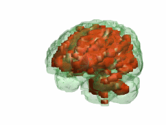
通过一次核磁共振扫描,首先从头部分割出脑体,然后确定脑容量。
Given an MRI scan, first segment the brain mass from the rest of the head, then determine the brain volume.
同时比较脑部存在的灰质和白质部分。
Also compare portions of gray and white matter present.
本示例是为研讨会而设计的。
This example was developed for seminars.
还用于2004年5月6日直播的医疗应用网络研讨会。
It was also used for webinars for medical applications broadcast live on May 6, 2004.
该软件包包括MATLAB代码和60幅DICOM图像的MRI扫描序列。
This package includes some MATLAB code and an MRI scan series consisting of 60 DICOM images.
完整源码下载地址:
http://page2.dfpan.com/fs/0lcc9jc222f10219163/
更多精彩文章请关注微信号: